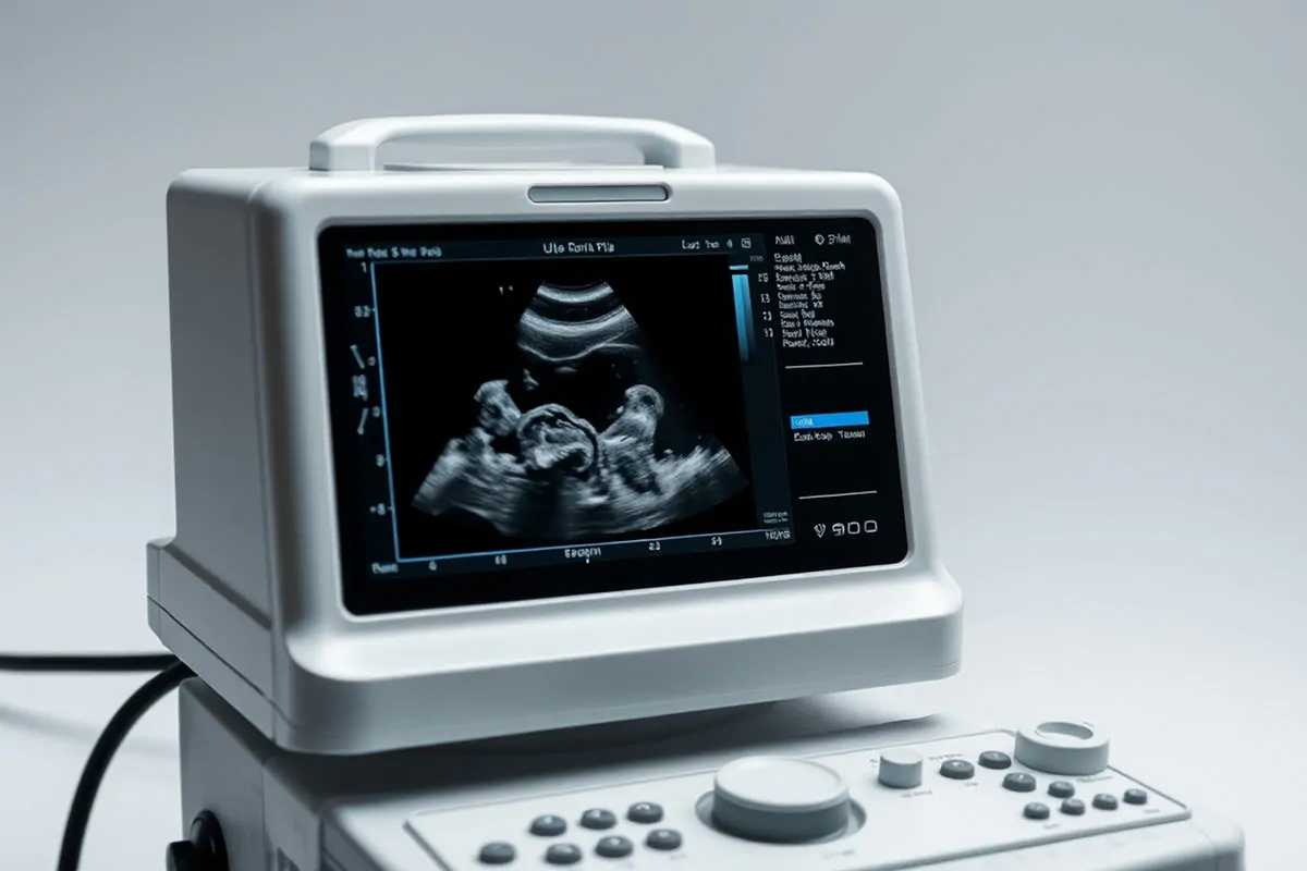
Ultrasonography

Ultrasonography is a medical imaging technique that uses high-frequency sound waves (ultrasound) to create images of internal organs and tissues.
During an ultrasound, a device called a transducer converts electrical energy into sound waves that are directed into the body. These sound waves bounce off structures and return to the transducer, which then converts them into electrical signals. A computer processes these signals to generate an image, which is displayed on a monitor and stored digitally. Since no X-rays are involved, ultrasonography does not expose the body to radiation.
Ultrasonography is a painless, cost-effective, and safe procedure, even during pregnancy.
Uses of Ultrasonography
Ultrasound provides real-time images, enabling the visualization of moving organs and structures, such as the beating heart, even in a fetus.
Ultrasonography is commonly used to detect growths or foreign objects near the body's surface, such as those found in the thyroid gland, breasts, testes, limbs, and some lymph nodes.
It is also used to examine internal organs in the abdomen, pelvis, and chest. However, sound waves cannot pass through gas (in the lungs or intestines) or bone, so specialized skills are needed for imaging internal organs. Sonographers, trained professionals, perform ultrasound examinations.
Ultrasonography is frequently used to evaluate:
- Heart: Detecting abnormalities in heart function, heart valve issues, and structural changes like enlargement (echocardiography).
- Blood vessels: Identifying dilation or narrowing of blood vessels.
- Gallbladder and biliary tract: Detecting gallstones and bile duct blockages.
- Liver, spleen, and pancreas: Identifying tumors or disorders.
- Urinary tract: Distinguishing benign cysts from potentially cancerous masses, and detecting kidney stones or blockages.
- Female reproductive organs: Identifying tumors, inflammation, or abnormalities in the ovaries, fallopian tubes, or uterus.
- Pregnancy: Monitoring fetal growth and development, and detecting placenta abnormalities, such as placenta previa.
- Musculoskeletal structures: Detecting joint swelling or ligament tears.
Ultrasonography can also assist doctors in guiding procedures like biopsies. By visualizing the target area and the biopsy instrument, it allows precise insertion and targeting.
